
 English
 French
 Pubmed
 Google Scholar
 Cross Ref
 Visistor: 1714456
A rare case of cavum tuberculosis
Un cas rare de tuberculose à cavum
S. Illé1, DA. Boubé2, ID. Bako3, N. Timi2, M. Ganda Aissa2, A. Dan Sono2
1: Service d’ORL et Chirurgie Cervico- Faciale de l’Hôpital Général de Référence de Niamey. Niger
2: Service d’ORL et Chirurgie Cervico- Faciale de l’Hôpital National de Niamey. Niger
3: Service de Radiologie de l’Hôpital Général de Référence de Niamey. Niger
Corresponding author:
Dr Illé Salha
Service d’ORL et Chirurgie Cervico- Faciale de l’Hôpital Général de Référence de Niamey. Niger
E-mail: ille_salha07@yahoo.fr
DOI: 10.12699/jfvpulm.12.36.2021.64
Introduction. Tuberculosis of the cavum is a rare localization of ENT disease. However, it remains a clinical reality that must be mentioned before any ENT pathology especially in the tropical context. Most often, it is a diagnostic surprise. The purpose of this work was to present a rare localization and clinical appearance of tuberculosis; to review the literature; and to specify the contribution of GeneXpert (PCR) in the diagnosis.
Case report. A 32-year-old patient who had been vaccinated with BCG since childhood, with no particular pathological history, was referred to us from a regional hospital for pharyngeal tumor. The ENT examination and the imaging (CT) were in favor of a retropharyngeal abscess, whose puncture confirmed the diagnosis by bringing back a purulent, yellowish, non-fetid liquid. Genxpert (PCR) from the same puncture fluid was used to isolate rifampicin-sensitive mycobacterium tuberculosis. The treatment was first surgical (incision-drainage of the purulent collection), then after the confirmation of the tuberculosis by the GeneXpert, a medical treatment antituberculosis was instituted during 6months. The evolution was favorable.
Conclusion. Tuberculosis of the cavum remains a rare manifestation of TB disease. We must think about any ENT pathology in a tropical environment. GeneXpert (PCR) was the decisive diagnostic argument.
KEYWORDS: Cavum tuberculosis; Retropharyngeal abscess; GeneXpert; Niger.
Introduction. La tuberculose du cavum est une localisation rare de la maladie ORL. Cependant, cela reste une réalité clinique qu'il faut évoquer avant toute pathologie ORL notamment dans le contexte tropical. Le plus souvent, c'est une surprise diagnostique. Le but de ce travail était de présenter une localisation rare et un aspect clinique de la tuberculose; passer en revue la littérature; et de préciser la contribution de GeneXpert (PCR) dans le diagnostic.
Rapport de cas. Un patient de 32 ans, vacciné au BCG depuis l'enfance, sans antécédent pathologique particulier, nous a été référé depuis un hôpital régional pour tumeur pharyngée. L'examen ORL et l'imagerie (TDM) étaient en faveur d'un abcès rétropharyngé, dont la ponction confirmait le diagnostic en rapportant un liquide purulent, jaunâtre et non fétide. Genxpert (PCR) à partir du même liquide de ponction a été utilisé pour isoler Mycobacterium tuberculosis sensible à la rifampicine. Le traitement a été d'abord chirurgical (incision-drainage de la collection purulente), puis après la confirmation de la tuberculose par le GeneXpert, un traitement médical antituberculeux a été instauré pendant 6 mois. L'évolution a été favorable.
Conclusion. La tuberculose du cavum reste une manifestation rare de la tuberculose. Il faut penser à toute pathologie ORL en milieu tropical. GeneXpert (PCR) a été l'argument diagnostique décisif.
MOTS CLÉS: Cavum tuberculosis; Abcès rétropharyngé; GeneXpert; Niger.
INTRODUCTION
ENT tuberculosis is a rare condition if lymph node locations are excluded [1]. Cavum tuberculosis represents less than 1% of the forms of this disease [2]. However , it remains a clinical reality that needs to be mentioned before any ENT pathology , particularly in the tropical context. Most often , it is a fortuitous discovery. Retropharyngeal abscess is also a rare but serious event that puts the patient’s life at risk. This gravity is explained by the risk of rupture and tracheobronchial flooding due to the absence of anatomical barrier between the cervical region and the mediastinum. We present here a rare localization and clinical aspect of tuberculosis ; to review the literature and to specify the diagnostic contribution of GeneXpert (PCR).
OBSERVATION
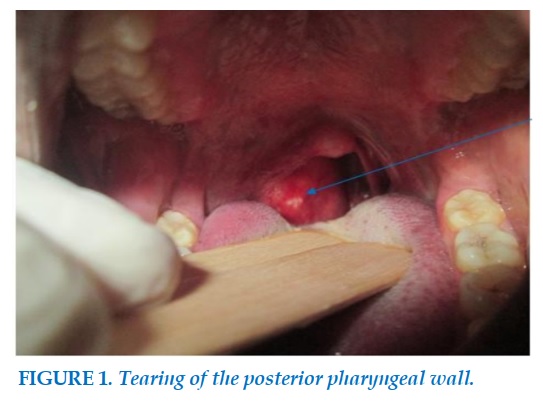
The patient was 32 years old , married, a shopkeeper, a rural resident; admitted to our ward for suspicion of a pharyngeal tumour. The onset of symptomatology would date back to about 2 months, with evening fever associated with high dysphagia , weight loss and hypersialorrhea. No odynophagia. Then the appearance of a non-inflammatory right lateral cervical swelling associated with rhinolalia and dyspnoea in the supine position. In this symptomatology, the patient consulted in a general hospital in the north of the country ,where the diagnosis of pharyngeal tumour was made and referred to the ENT department and cervico-facial surgery of the National Hospital of Niamey, for specialized care. In his antecedents , there was a notion of herbal treatment and taking Amoxicillin clavulanic acid for 10 days, without improvement. In view of the persistence of the above clinical signs , this patient was referred to our department. On physical examination, the patient had a good general condition, a temperature of 38,5°C. The oropharyngoscopy objectified, a curvature of the posterior pharyngeal wall, painless, clean, non-pulsatile, more prominent in the right peripharyngeal (Figure 1).
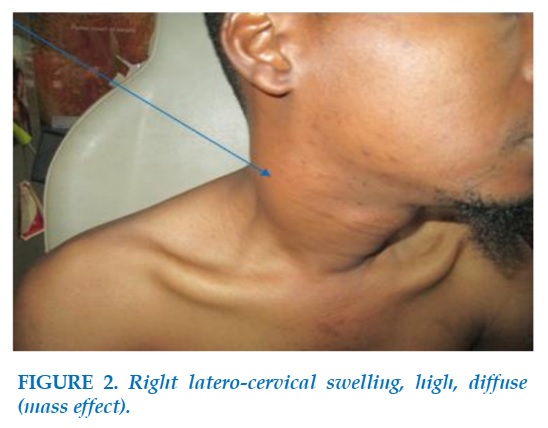
The palatine tonsils, the veil are without particularity. Cervical examination highlights, right lateral cervical swelling (Figure 2), high, diffuse ,due to a mass effect of pharyngeal swelling on the latero-cervical soft tissues. No palpable cervical lymphadenopathy. The rest of the ENT clinical examination was unremarkable .
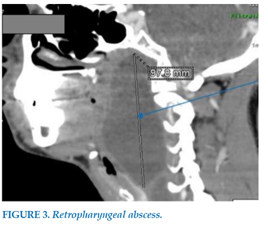
Examination of other devices is normal. The pharyngeal scan, highlighted, a retro- pharyngeal abscess (Figure 3), whose puncture confirmed the diagnosis, by bringing back a purulent, yellowish, non -fetid liquid.
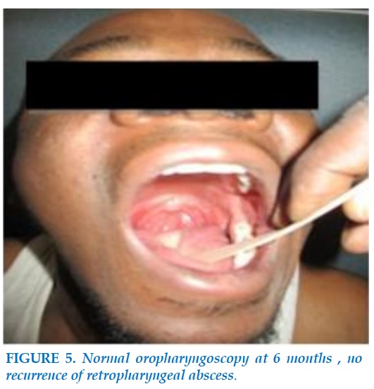
The biological assessment realized , in particular, the blood count was normal, the sedimentation rate at 45 mm in the first hour, Blood glucose at 6.1 mmol/l. HIV1 and 2 serology was negative. The tuberculin IDR was 20 mm. GeneXpert on the pharyngeal puncture fluid was positive with detection of mycobacterium tuberculosis susceptible to rifampicin. The chest x-ray was normal . As part of a multidisciplinary care, an opinion from the infectious disease specialist was requested after confirmation of TBC by GeneXpert for TB treatment. Before the initiation of the antituberculous medical treatment, an incision-drainage of the abscess (figure4) was performed at the operating block, under general anaesthesia. The anti-tuberculosis treatment RHZE regimen (rifampicin, isoniazid, pyrazinamide, ethambutol) 2 months, RH 4 months was instituted with a good clinical evolution. The operative sequences were simple. The evolution was favourable after 6 months of treatment without recurrence (Figure 5).
DISCUSSION
Nasopharyngeal tuberculosis is a rare localization of the disease in its primitive form, although the cavum is richly vascularized and located in a very widely exposed area [3,4]. This rarity found in our service ,is also reported in the literature [5,6]. As is the case with our patient , the tuberculosis of the cavum mainly affects the young adult between 20 and 40 years (extremely 8 and 62 years) ; and his clinical signs are not specific [2]. The predominance of extra-pulmonary tuberculosis in adults may be explained by the fact that the efficacy of BCG vaccination is not absolute [7]. The effectiveness of BCG vaccination is limited to the protection against the deadly evolution of tuberculosis , especially tuberculous meningitis and disseminated disease (miliary) [7]. It does not prevent the transmission of the disease and halt the global epidemic . The vaccine is more effective in new-born and child than in adult protection estimated between 75 and 85 % of serious forms of infants and young children and between 50 et 75 % of the forms of the adult [7]. Moreover , it must be remembered that tuberculosis is very confusing and can present itself in various macroscopic aspects. It most often assumes pseudo-tumoral forms [8] posing the differential diagnosis with a tumour of the cavum. The retropharyngeal abscess is exceptional in the adult [9], likewise, its tubercular origin is however not usual. Clinically , the conjunction of generally unilateral rhinologic signs , associated with evening fever as in this patient , are the most common modes of revelation.
The median or paramedian arch of the posterior pharyngeal wall is almost constant in the retropharyngeal abscess [9]. Pharyngeal obstruction may be the cause of dysphagia , dyspnoea and rhinolalia. Advances in biology have led to fairly sensitives diagnoses in shorter time frames.
In resource–poor countries where it is a real public health problem, of TB is often a problem because of limited technical expertise and the inconstancy with which the various confirmatory examinations are carried out. The introduction of new tools, essentially of molecular biology including Xpert/MTB/ Rif or GeneXpert® test whose use has been approved since December 2010 by WHO ; has increased sensitivity and especially to shorten the time of confirmation of tuberculosis [10]. This test is now available and usable in Niger, and our study reports one of its first experiences of use.
GeneXpert has been of great benefit for the early detection of pulmonary and extra-pulmonary tuberculosis but also for the detection of resistance to rifampicin, problem of growing concern, especially in a context where culture was not available routinely [11]. GeneXpert can provide formal proof within a reasonable time (less than 2 hours), unlike bacteriology whose result (culture) may require several months [11].
However , in the diagnosis of tuberculosis , classical examinations such as intradermoreaction to tuberculin , sedimentation rate, la radiology, histological examination, remain the reference approach and should remain essential in the management of patients suspected of having tuberculosis[12]. The treatment of tuberculosis of the cavum is medical [2]. It is based on anti-TB drugs in combination over a prolonged period. It is subject to an obligation of prior diagnosis (bacteriology or histology). It usually associates Rifampicin (10mg/kg/j), Isoniazid (5mg/kg/j), Ethambutol (15 mg/kg/j) And Pyrazinamide (20 à 30 mg/kg/j) for 2 months then rifampicin, pyrazinamide for 4 months ( RHZ-2/RH- 4).Surgery has limited. In our case, the compressive risk of the upper aerodigestive tract by the large volume of the retropharyngeal abscess, but especially the risk of rupture and flood tracheobronchial or the occurrence of formidable complications such as mediastinitis and Grisel’syndrome, justified the incision-drainage performed in this patient. The evolution was 6 months without recurrence; testifying to our adequate therapy in this patient. The risk of relapse is estimated at 1%,mainly due to the appearance of multi-resistant BK strains [8]; which was not the case in our patient.
CONCLUSION
This clinical case confirms the clinical expression of cavum tuberculosis in the form of retropharyngeal abscess. The GeneXpert shows its definite contribution in the diagnosis of confirmation of tuberculosis.
CONFLICT OF INTERESTS
Non.
REFERENCES
1. El Ayoubi A, Benhammou A, El Ayoubi F, El Fahssi A, Nitassi S, Kohen A, et al. La tuberculose primitive ORL extra ganglionnaire Annales d'Otolaryngologie et de Chirurgie Cervico-faciale2009, 126,(4):208-15.
2. Bouaity B, Nadour k, Al Jalil A, Touihem N, Attifi H, Kettani M et coll. La tuberculose du cavum. La Lettre d’oto-rhino-laryngologie et de chirurgie cervico-faciale 2009;319:14-16.
3. Tse GM, Ma TK, Chan AB, et al. Tuberculosis of the nasopharynx: a rare entity revisited. Laryngoscope. 2003 Apr;113(4):737–40.
4. Gassab E, Kedous S, Berkaoui A, Sayeh N, Harrathi K, Koubaa J, Gassab A. Tuberculose extra ganglionnaire de la tête et du cou J. Tun Orl 2010,(24) : 26-29.
5. Ito K, Morooka M, Kubota K. Findings of pharyngeal tuberculosis. Ann Nucl Med 2010; 24:493-6.
6. Kuran G, Sagit M, Saka C et al. Nasopharyngeal tuberculosis: an unusual cause of nasal obstruction and snoring. B-ENT 2008;4:249-51.
7. J Antoine D, Guthmann JP, LEVY-BRUHL D, CHE D. Impact des modifications des modalités de vaccination par le BCG sur l’épidémiologie de la tuberculose en France en 2009. BEH 2011;22:255–7.
8. Mohammed T, Abdelfettah A, Mehdi C, Rachid B, Brahim B et al. Tuberculose primitive du cavum d’aspect pseudo tumoral. Pan Afr Med J 2013,14 :63-67.
9. Benmansour et coll. Abcès rétropharyngé chez l’adulte. Rev Laryngol Otol Rhino 2012; 133(3):137-139.
10. Ninet B, Roux-Lombard P, Schrenzel J, Janssens J. New tests for the diagnosis of tuberculosis. Rev Mal Respir2011; 28:823-33.
11. Blanie M, Pellegrin J, Maugein J. Apport de la PCR dans le diagnostic des tuberculoses extrapulmonaire. Médecine et maladies infectieuses 2005;35:17- 22.
12. Sylvie A D, Aminata M, Daye K, Noel M M, Louise FD, Cheikh Tidiane N et coll. Utilisation du test GeneXpert pour le diagnostic de la tuberculose au service des maladies infectieuses du CHNU de Fann. The Pan African Medical Journal 2016; 26:23-244.

-FIGURES-
REFERENCES
1. El Ayoubi A, Benhammou A, El Ayoubi F, El Fahssi A, Nitassi S, Kohen A, et al. La tuberculose primitive ORL extra ganglionnaire Annales d'Otolaryngologie et de Chirurgie Cervico-faciale2009, 126,(4):208-15.
2. Bouaity B, Nadour k, Al Jalil A, Touihem N, Attifi H, Kettani M et coll. La tuberculose du cavum. La Lettre d’oto-rhino-laryngologie et de chirurgie cervico-faciale 2009;319:14-16.
3. Tse GM, Ma TK, Chan AB, et al. Tuberculosis of the nasopharynx: a rare entity revisited. Laryngoscope. 2003 Apr;113(4):737–40.
4. Gassab E, Kedous S, Berkaoui A, Sayeh N, Harrathi K, Koubaa J, Gassab A. Tuberculose extra ganglionnaire de la tête et du cou J. Tun Orl 2010,(24) : 26-29.
5. Ito K, Morooka M, Kubota K. Findings of pharyngeal tuberculosis. Ann Nucl Med 2010; 24:493-6.
6. Kuran G, Sagit M, Saka C et al. Nasopharyngeal tuberculosis: an unusual cause of nasal obstruction and snoring. B-ENT 2008;4:249-51.
7. J Antoine D, Guthmann JP, LEVY-BRUHL D, CHE D. Impact des modifications des modalités de vaccination par le BCG sur l’épidémiologie de la tuberculose en France en 2009. BEH 2011;22:255–7.
8. Mohammed T, Abdelfettah A, Mehdi C, Rachid B, Brahim B et al. Tuberculose primitive du cavum d’aspect pseudo tumoral. Pan Afr Med J 2013,14 :63-67.
9. Benmansour et coll. Abcès rétropharyngé chez l’adulte. Rev Laryngol Otol Rhino 2012; 133(3):137-139.
10. Ninet B, Roux-Lombard P, Schrenzel J, Janssens J. New tests for the diagnosis of tuberculosis. Rev Mal Respir2011; 28:823-33.
11. Blanie M, Pellegrin J, Maugein J. Apport de la PCR dans le diagnostic des tuberculoses extrapulmonaire. Médecine et maladies infectieuses 2005;35:17- 22.
12. Sylvie A D, Aminata M, Daye K, Noel M M, Louise FD, Cheikh Tidiane N et coll. Utilisation du test GeneXpert pour le diagnostic de la tuberculose au service des maladies infectieuses du CHNU de Fann. The Pan African Medical Journal 2016; 26:23-244.
ARTICLE INFO
DOI: 10.12699/jfvpulm.12.36.2021.56
Conflict of Interest
Non
Date of manuscript receiving
15/12/2020
Date of publication after correction
15/01/2021
Article citation
Illé S., Boubé DA., Bako ID., Timi N., Ganda Aissa M., Dan Sono A. A rare case of cavum tuberculosis. J Func Vent Pulm 2021;36(12):64-67


Copyright: jfvpulm.com