
 English
 French
 Pubmed
 Google Scholar
 Cross Ref
 Visistor: 1968648
Drug-Induced Sleep Endoscopy (DISE) with Real-Time Sleep EEG Monitoring Using Flexible Nasopharyngoscopy at Quang Ninh Provincial General Hospital, Vietnam
Endoscopie sous sommeil induit (DISE) avec monitorage EEG en temps réel par nasopharyngoscopie flexible au sein de l'Hôpital général provincial de Quang Ninh, Vietnam
Vu Thanh Khoa¹, Pham Van Hieu¹, Bui Thanh Huong¹, Pham Quang Huy 1, Dinh Thi Hue¹, Nguyen Thi Ngan¹, Do Huyen Trang¹, Nguyen Ba Viet¹, Sy Duong-Quy¹,²
1Quang Ninh General Hospital, Quang Ninh, Vietnam.
2President, Vietnam Society of Sleep Medicine - VSSM
Corresponding author:
Sy Duong-Quy. Sleep Medicine Research Center. Vietnam Society of Sleep Medicine. Lam Dong, Viet Nam.
Email: :sduongquy.jfvp@gmail.com
ABSTRACT
Obstructive Sleep Apnea (OSA) is one of the most common sleep-related breathing disorders, characterized by recurrent collapse of the upper airway during sleep, resulting in intermittent hypoxemia, sleep fragmentation, and disruption of normal sleep architecture. The pathophysiology of OSA is multifactorial and may involve reduced pharyngeal muscle tone, abnormalities in central neural control, and structural alterations of the upper airway. Structural upper airway abnormalities represent a major contributing factor, particularly among younger patients.
Polysomnography (PSG) remains the gold standard for diagnosing OSA and determining disease severity using the Apnea–Hypopnea Index (AHI). However, PSG cannot accurately identify the anatomical site or pattern of upper airway obstruction. Drug-Induced Sleep Endoscopy (DISE) enables dynamic assessment of the upper airway under a condition closely resembling physiological sleep, allowing precise identification of the location, pattern, and degree of obstruction. This information facilitates individualized treatment planning and surgical decision-making.
The use of DISE in clinical practice remains limited in Vietnam. This report presents DISE procedures integrated with real-time electroencephalographic (EEG) monitoring using the FrenzBand device for pre- and post-operative assessment of upper airway obstruction, highlighting the value of DISE within a multidisciplinary sleep medicine approach.
KEYWORDS: DISE; Obstructive Sleep Apnea; OSA; Sleep Endoscopy; VOTE Classification; Upper Airway.
RÉSUMÉ
Le syndrome d'apnées obstructives du sommeil (SAOS) est l'un des troubles respiratoires liés au sommeil les plus fréquents. Il se caractérise par un collapsus récurrent des voies aériennes supérieures pendant le sommeil, entraînant une hypoxémie intermittente, une fragmentation du sommeil et une altération de l'architecture normale du sommeil. La physiopathologie du SAOS est multifactorielle et peut impliquer une réduction du tonus des muscles pharyngés, des anomalies du contrôle neural central et des altérations structurelles des voies aériennes supérieures. Ces anomalies structurelles constituent un facteur contributif majeur, particulièrement chez les patients jeunes.
La polysomnographie (PSG) demeure la référence absolue (gold standard) pour diagnostiquer le SAOS et déterminer la sévérité de la maladie par l'indice d'apnées-hypopnées (IAH). Cependant, la PSG ne permet pas d'identifier avec précision le siège anatomique ou le schéma de l'obstruction des voies aériennes supérieures. L'endoscopie sous sommeil induit (DISE) permet une évaluation dynamique des voies aériennes supérieures dans des conditions proches du sommeil physiologique, autorisant une identification précise de la localisation, du schéma et du degré d'obstruction. Ces informations facilitent la planification thérapeutique individualisée et la prise de décision chirurgicale.
L'utilisation de la DISE en pratique clinique reste limitée au Vietnam. Ce rapport présente des procédures de DISE intégrées à un monitorage électroencéphalographique (EEG) en temps réel, utilisant le dispositif FrenzBand, pour l'évaluation pré et post-opératoire de l'obstruction des voies aériennes supérieures, soulignant ainsi la valeur de la DISE dans une approche multidisciplinaire de la médecine du sommeil.
MOTS-CLÉS: DISE; SAOS; Endoscopie du sommeil; Classification VOTE; Voies aériennes supérieures.
Objectives
To describe the technical procedure of Drug-Induced Sleep Endoscopy (DISE) using a flexible endoscope and to evaluate its role in assessing the site, pattern, and severity of upper airway obstruction using the VOTE classification in patients with suspected or diagnosed Obstructive Sleep Apnea (OSA).
Subjects and Methods
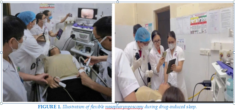
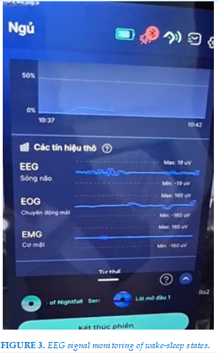
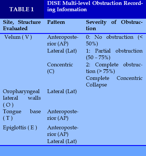
Clinical cases included patients with suspected or diagnosed OSA at Quang Ninh General Hospital who required pre-treatment evaluation of obstruction sites or post-operative assessment following upper airway obstruction intervention. Patients fasted for 6–8 hours prior to the procedure; vital signs and $SpO_2$ were continuously monitored. Intravenous sedation was induced using propofol at a mean concentration of 3.2 µg/mL, with sleep onset verified by EEG monitoring using the Frenzband device. Once the patient reached a sleep state and snoring occurred, flexible nasopharyngoscopy was performed using a 2.7 mm Pentax endoscope. The sites surveyed included the Velum, Oropharynx, Tongue base, and Epiglottis. Obstruction was evaluated based on the VOTE classification (site, pattern, and severity) (Figure 1). A chin-lift maneuver was combined to assess changes in oxygen saturation and potential airway improvement.
Classification Criteria (Table 1):
Severity:
Pattern:
Site:
Results
DISE allows for direct and dynamic visualization of upper airway collapse sites during 2–3 snoring cycles at each anatomical level. This method enables the identification of associated abnormalities, such as septal deviation, hypertrophic turbinates, adenoid hypertrophy, palatine/lingual tonsillar hypertrophy, lateral pharyngeal wall collapse, and epiglottic collapse.
Furthermore, DISE facilitates the immediate assessment of the chin-lift maneuver's effectiveness on airway obstruction and SpO2. The procedure was performed safely through the collaboration of Otolaryngologists and Anesthesiologists, with patients recovering stably under observation.
Clinical Case
Case Report 1
A 34-year-old male patient was admitted to the hospital due to snoring. Family members reported that the patient snored loudly and experienced excessive daytime sleepiness.
Otolaryngologic endoscopic examination: The nasal passages were clear, the turbinates were normal, and no anatomical deformities were observed within the nasal cavity. The nasopharyngeal vault appeared smooth; both palatine tonsils were Grade 2, and the uvula was normal. There was minimal lymphoid tissue at the base of the tongue, and the epiglottis was normal.
Clinical Scores:
Polysomnography (PSG) results:
Diagnosis: Severe Obstructive Sleep Apnea (OSA) and chronic tonsillitis.
The patient was indicated for surgery, including tonsillectomy, uvulopalatopharyngoplasty (UPPP) with total uvulectomy, and bilateral pillar suturing.
One month post-operatively, the patient underwent DISE at Quang Ninh General Hospital, with intravenous propofol sedation and upper airway assessment monitored by the FrenzBand device to verify sleep state.
Clinically, the patient no longer experienced snoring and did not report the morning fatigue felt prior to the surgery. Flexible nasopharyngoscopy was performed using a 2.7 mm Pentax endoscope (Figure 1). Throughout the procedure, the patient was monitored via real-time EEG using the FrenzBand to ensure sleep status and optimize sedation levels (Figure 2). Obstruction was assessed according to the VOTE classification.
DISE findings indicated:
Total hypopharyngeal narrowing score: 1 point.
Case Report 2 A 16-year-old male patient presented with complaints of snoring. Family members reported loud snoring and a sensation of lump in the throat (globus pharyngeus).
Otolaryngologic endoscopic examination: The nasal passages contained small amounts of clear secretions, the turbinates were slightly enlarged and erythematous, with no anatomical deformities within the nasal cavity. Adenoid tissue was Grade 2; both palatine tonsils were Grade 2, the uvula appeared normal, and the epiglottis was normal.
Clinical Scores:
Diagnosis: Rhinitis and chronic tonsillitis.
Flexible nasopharyngoscopy was performed using a 2.7 mm Pentax endoscope. Throughout the procedure, the patient was monitored via real-time EEG using the FrenzBand device to verify sleep state and optimize sedation levels. Obstruction was assessed according to the VOTE classification.
DISE findings indicated:
The patient was advised to undergo polygraphy (respiratory sleep study) prior to surgery for tonsillectomy and uvulopalatopharyngoplasty (Figure 2).
Discussion
For Patient 1, the DISE results yielded a total score of 1, indicating that the UPPP surgery successfully resolved obstruction in the velum, uvula, and lateral pharyngeal walls; however, the patient still exhibited incomplete tongue base collapse. The patient's clinical outcome was considered good, with no further indication for polysomnography.
For Patient 2, the initial endoscopic examination showed Grade 2 tonsils; however, during drug-induced sleep, a bilateral narrowing equivalent to Grade 4 tonsillar hypertrophy was observed. Combined with incomplete concentric narrowing of the velum and tongue base, the patient was assessed as having a high risk of obstructive sleep apnea (OSA) and was advised to undergo a respiratory sleep study prior to any surgical intervention.
The post-procedure recovery time for patients ranged from 6 to 10 minutes, and patients were fully alert and ready for discharge within 30 minutes.
DISE results revealed multi-level obstruction, primarily at the level of the velum and oropharynx. DISE plays a crucial role in the individualization of treatment. Rather than relying solely on the Apnea–Hypopnea Index (AHI), DISE assists in precisely identifying the location and pattern of airway collapse, thereby supporting the selection of an optimal treatment strategy.
A highlight of these cases is the application of real- time EEG monitoring using the FrenzBand
during DISE. EEG monitoring helps confirm a physiological-like sleep state, avoiding excessive sedation—a factor that could potentially skew results. This technique contributes to more accurate and objective assessment and surgical decision-making.
This report also demonstrates the development potential of the sleep medicine and airway endoscopy specialty in Vietnam, particularly through a multidisciplinary approach involving Otolaryngology, Anesthesiology, Neurology, and Sleep Medicine.
Conclusion
Drug-induced sleep endoscopy using a flexible endoscope is a safe and effective method for evaluating
the dynamics of the upper airway in patients with OSA. This technique helps precisely identify the site and pattern of obstruction according to the VOTE classification, thereby supporting the selection of appropriate and personalized treatment methods for each patient.
These cases represent the first instances in Vietnam where the DISE procedure has been integrated with real-time EEG monitoring using the FrenzBand device to assess patients before and after OSA intervention. This methodology enhances the reliability of endoscopic assessment and supports the optimization of treatment strategies for complex OSA cases.
REFERENCES
| 1. Kezirian EJ, et al. Drug-Induced Sleep Endoscopy: Updated International Consensus. Sleep Med Clin. 2024. |
| 2. Huntley C, et al. DISE and Personalized OSA Surgery. Otolaryngol Clin North Am. 2023. |
| 3. Ravesloot MJL, et al. Advances in DISE Classification Systems. Sleep Breath. 2022. |
| 4. Stefani A, et al. Sleep Disorders in Parkinson Disease. Lancet Neurol. 2023. |
| 5. Arnulf I. Parkinson Disease and REM Sleep Behavior Disorder. Neurology. 2022. |
| 6. Vicini C, et al. DISE in Multilevel OSA Surgery. Eur Arch Otorhinolaryngol. 2021. |
| 7. American Academy of Sleep Medicine Clinical Updates. 2024. |
| 8. ASOHNS/ASHNO educational updates on sleep surgery and DISE. 2023–2025. |
| 9. Duong-Quy, S., Chirakalwasan, N., Nguyen-Nhu, V., & Craig, T. Sleep disorders and airway diseases. Frontiers in Neurology 2023, 14, 1264665. |
| 10. Nguyen, A., Pogoncheff, G., Dong, B. X., Bui, N., Truong, H., Pham, N., DuongQuy,S, ... & Vu, T. A comprehensive study on the efficacy of a wearable sleep aid device featuring closed-loop real-time acoustic stimulation. Scientific Reports, 13(1), 17515. |
| 11. Duong-Quy, S., Nguyen-Huu, H., Hoang-Chau-Bao, D., Tran-Duc, S., Nguyen-ThiHong, L., Nguyen-Duy, T., ... & Craig, T. Personalized medicine and obstructive sleep apnea. Journal of Personalized Medicine 2022, 12(12), 2034. |
| 12. Diep, L. V., Trang, N. M., Phap, T. Q., Chung, T. N., Bo, N. T. M., Xuan, N. T., Tri, N. M., Long, H., Tuyen, H. T., & Sy, D. Q. (2026). Drug Induced Sleep Endoscopy of the Upper Airway at 199 Hospital. Journal Francophone of Ventilation - Pulmonology and Sleep, 2026. |

FIGURES - TABLES
REFERENCES
| 1. Kezirian EJ, et al. Drug-Induced Sleep Endoscopy: Updated International Consensus. Sleep Med Clin. 2024. |
| 2. Huntley C, et al. DISE and Personalized OSA Surgery. Otolaryngol Clin North Am. 2023. |
| 3. Ravesloot MJL, et al. Advances in DISE Classification Systems. Sleep Breath. 2022. |
| 4. Stefani A, et al. Sleep Disorders in Parkinson Disease. Lancet Neurol. 2023. |
| 5. Arnulf I. Parkinson Disease and REM Sleep Behavior Disorder. Neurology. 2022. |
| 6. Vicini C, et al. DISE in Multilevel OSA Surgery. Eur Arch Otorhinolaryngol. 2021. |
| 7. American Academy of Sleep Medicine Clinical Updates. 2024. |
| 8. ASOHNS/ASHNO educational updates on sleep surgery and DISE. 2023–2025. |
| 9. Duong-Quy, S., Chirakalwasan, N., Nguyen-Nhu, V., & Craig, T. Sleep disorders and airway diseases. Frontiers in Neurology 2023, 14, 1264665. |
| 10. Nguyen, A., Pogoncheff, G., Dong, B. X., Bui, N., Truong, H., Pham, N., DuongQuy,S, ... & Vu, T. A comprehensive study on the efficacy of a wearable sleep aid device featuring closed-loop real-time acoustic stimulation. Scientific Reports, 13(1), 17515. |
| 11. Duong-Quy, S., Nguyen-Huu, H., Hoang-Chau-Bao, D., Tran-Duc, S., Nguyen-ThiHong, L., Nguyen-Duy, T., ... & Craig, T. Personalized medicine and obstructive sleep apnea. Journal of Personalized Medicine 2022, 12(12), 2034. |
| 12. Diep, L. V., Trang, N. M., Phap, T. Q., Chung, T. N., Bo, N. T. M., Xuan, N. T., Tri, N. M., Long, H., Tuyen, H. T., & Sy, D. Q. (2026). Drug Induced Sleep Endoscopy of the Upper Airway at 199 Hospital. Journal Francophone of Ventilation - Pulmonology and Sleep, 2026. |
ARTICLE INFO DOI:10.12699/jfvpulm.17.53.2026.53
Conflict of Interest
Non
Date of manuscript receiving
10/03/2025
Date of publication after correction
26/06/2026
Article citation
Vu Thanh Khoa, Pham Van Hieu, Bui Thanh Huong, Pham Quang Huy, Dinh Thi Hue, Nguyen Thi Ngan, Do Huyen Trang, Nguyen Ba Viet, Sy Duong-Quy. Drug-Induced Sleep Endoscopy (DISE) with Real-Time Sleep EEG Monitoring Using Flexible Nasopharyngoscopy at Quang Ninh Provincial General Hospital, Vietnam. J Fran Vent PulmS 2026;53(17):53-56